Genes that are part of 22 pairs of non-sex chromosomes are called autosomal. Autosomal dominant diseases are diseases in which one mutant gene (allele) in a heterozygous state is sufficient for the appearance of phenotypic manifestations.
Autosomal dominant inheritance of diseases is characterized by a number of the following signs, which are detected in most cases:
- the disease is transmitted vertically along the pedigree, and cases of the disease are diagnosed in each generation;
- the risk of inheriting the disease for any of the patient's children is 50%;
- phenotypically normal family members do not inherit diseases to their offspring;
- both sexes are affected with equal frequency;
- a significant proportion of cases of the disease are caused by a new mutation.
With autosomal dominant inheritance, in contrast to the X-linked type, transmission of the disease through the male line is possible. Since a man passes on a Y rather than an X chromosome to his sons, in cases where a hereditary disease is passed from father to son, X-linked inheritance is excluded.
With autosomal dominant inheritance, there is significant variability in clinical manifestations within the family. In most cases, this is due to variable expression of the mutant gene. The exact cause of this variability is unknown, but it is most likely that it is due to the influence of modifier genes and environmental factors on the phenotype. In some families, obligate carriers of the mutant gene do not have phenotypic manifestations of the disease. This phenomenon of autosomal dominant inheritance is called incomplete penetrance, i.e. inheritance follows the “all or nothing” principle. In some cases, when there is an impression of a lack of penetrance, the patient may have a low degree of somatic mosaicism or germline cell mosaicism for this gene. Somatic mosaicism occurs at the stage of embryonic development due to a mutation in a somatic cell, leading to the formation of mixed genotypes in fetal cells, some of which contain the mutation, while others lack it. In typical cases, the effects of the mutant gene are less pronounced or absent in these patients. Germline cell mosaicism occurs in the embryo after conception and is limited to cells that are the precursors of eggs or sperm. It is often observed in conditions such as osteogenesis imperfecta and syndromes associated with craniostenosis (Apert and Crouzon syndromes).
Since a single mutant gene is sufficient for the phenotypic manifestations of autosomal dominant inheritance, these conditions arise in many patients as a result of a new mutation. The more severe the disease, the greater the incidence of cases arising from de novo gene mutations. In severe diseases, decreased reproductive function limits the transmission of the mutant gene. In some cases of the occurrence of a new mutation, the parents are elderly (over 40 years old).
The article was prepared and edited by: surgeonVideo:
Healthy:
Related articles:
- Theoretically, individuals carrying a dominant trait can be homozygous or heterozygous. However, when it comes to...
- Autosomal recessive diseases include diseases in which two copies of a mutant are required for the appearance of phenotypic characteristics...
The most typical diseases with an autosomal recessive type of inheritance are cystic fibrosis, phenylketonuria, galactosemia, adrenogenital syndrome, and mucopolysaccharidosis. The peculiarities of sex-linked diseases are due to the fact that women have two X chromosomes, and men have one. A woman receives her two X chromosomes and corresponding genes from both her father and mother, while a man inherits his only X chromosome only from his mother. A woman, having inherited a pathological gene from one of her parents, is heterozygous, and a man is hemizygous, since genes located on the X chromosome do not have alleles on the Y chromosome. In this regard, traits inherited in an X-linked manner occur in a population with different probabilities in males and females. Inheritance linked to sex chromosomes can be dominant or recessive (usually recessive).
The type of inheritance is autosomal dominant. types of inheritance of traits in humans
Some dominant genetic diseases appear immediately after birth. Others may not appear until adulthood; such diseases are called “late-onset diseases” or “late-onset diseases.”
Attention
Examples of such diseases are polycystic kidney disease in adults and Huntington's chorea. How are dominant diseases inherited? Figure 2: How dominant diseases are transmitted from parent to child If one of the parents has an altered copy of the gene, then he can pass on either the normal copy or the altered copy to the child.
Thus, each of such a parent's children would have a 50% chance of inheriting the altered copy and therefore having the genetic disease.
Autosomal dominant type of inheritance
Normally, hemizygous genes are those localized on the sex chromosomes of the heterogametic sex, i.e., the sex that produces different types of germ cells. Hemizygosity also occurs as a result of aneuploidy or deletion, when only one of a pair of allelic genes is retained in the genotype, which can manifest itself as a recessive mutation.
Diseases characterized by X-linked dominant inheritance include vitamin D-resistant rickets (rickets that cannot be treated with regular doses of vitamin D), orofacial-digital syndrome (multiple hyperplastic frenulum of the tongue, cleft lip and palate, alar hypoplasia nose, asymmetrical shortening of fingers) and other diseases.
Dominant inheritance
At a low level of penentrance, the mutant gene may not appear in every generation. Most often, the type of inheritance is autosomal dominant, which transmits diseases from generation to generation.
With this type of inheritance in a sick child, one of the parents suffers from the same disease. However, if only one parent in a family is sick, and the other has healthy genes, then the children may not inherit the mutant gene.
An example of inheritance according to an autosomal dominant type. The type of autosomal dominant inheritance can transmit more than 500 different pathologies, among them: Marfan syndrome, Ehlers-Danlos syndrome, dystrophy, Recklinghuisen disease, Huntington disease. When studying the pedigree, one can trace the autosomal dominant type of inheritance.
There may be different examples of this, but the most striking is Huntington’s disease. It is characterized by pathological changes in nerve cells in the structures of the forebrain.
Important
Mosaicism is observed in many chromosomal diseases. It is believed that somatic mutations and mosaicism play an important role in the etiology of many types of malignant neoplasms.
Mosaicism also occurs among germ cells. During oogenesis, 28-30 mitotic divisions occur, and during spermatogenesis - up to several hundred. In this regard, with non-somatic mosaicism, the frequency of manifestation of the mutation and the risk of its transmission to next generations increase.Nonsomatic mosaicism is observed in osteogenesis imperfecta and some diseases inherited linked to the X chromosome. 2. Mitochondrial diseases. Mitochondria have their own DNA; mtDNA is located in the matrix of the organelle and is represented by a circular chromosome.
It is believed that during cell division, mitochondria are randomly distributed between daughter cells.
Criteria for different types of inheritance
A. Monogenic inheritance. A trait encoded by one gene is inherited in accordance with Mendel's laws and is called Mendelian. The totality of all the genes of an organism is called the genotype.
A phenotype is the realization of a genotype (in morphological and biochemical terms) under specific environmental conditions. 1. One of the possible structural states of a gene is called an allele.
Alleles arise as a result of mutations. The potential number of alleles for each gene is practically unlimited. In diploid organisms, a gene can be represented by only two alleles, localized on identical sections of homologous chromosomes.
The condition when homologous chromosomes carry different alleles of the same gene is called heterozygous. 2. Inheritance of monogenic diseases - autosomal or X-linked - can be determined by studying the pedigree.
Types of inheritance of diseases
Autosomal dominant diseases are characterized by a number of the following signs, which are detected in most cases: 1) the disease is transmitted vertically along the pedigree, and cases of the disease are diagnosed in each generation, 2) the risk of inheriting the disease for any of the patient’s children is 50%; 3) phenotypically normal family members do not inherit diseases to their offspring; 4) males and females are affected with equal frequency: 5) a significant proportion of cases of the disease are caused by a new mutation. With an autosomal dominant type of inheritance, in contrast to the X-linked type, transmission of the disease through the male line (from father to son) is possible (Fig.
29.2). Since a man passes on a Y rather than an X chromosome to his sons, in cases where a hereditary disease is passed from father to son, X-linked inheritance is excluded.
News of website sections
Healthy family members give birth to healthy children. 4) Autosomal dominant diseases are always inherited, regardless of the sex of the child and the sex of the affected parent. Exceptions occur in cases of new mutations and incomplete gene penetrance.
b. Autosomal recessive inheritance. Diseases with an autosomal recessive pattern of inheritance include Tay-Sachs disease, cystic fibrosis, and most inherited metabolic disorders. Autosomal recessive diseases are usually more severe than autosomal dominant diseases.
1) If both parents are healthy but are carriers of a pathological gene, the risk of having a sick child is 25%. 2) A healthy child in 2/3 of cases turns out to be a heterozygous carrier of the pathological gene. 3) In a child with an autosomal recessive disease, especially a rare one, the parents often turn out to be blood relatives.
Forbidden
Since in a sick parent the mutant gene is localized in half of the gametes, which can be fertilized equally with normal cells, the probability of the disease occurring in children is 50%. However, when analyzing pedigrees, it is necessary to remember the possibility of incomplete penetration of the dominant allele due to the interaction of genes or environmental factors.
All phenotypically healthy children will be genetically healthy if the penetrance of the mutant gene is complete. In cases of low penetrance, pathological signs do not appear in some generations.
It should also be noted that some diseases do not appear from the moment of birth, but only at a certain age. This creates certain difficulties in establishing the type of inheritance.
The autosomal recessive type of inheritance of the trait is transmitted
An example of an X-linked recessive disease is hemophilia A, characterized by a blood clotting disorder due to deficiency of factor VIII - antihemophilic globulin A. The pedigree of a patient with hemophilia is shown in Fig. IX. 11. Clinically, the disease is manifested by frequent prolonged bleeding even with a minor wound, hemorrhages into organs and tissues. The incidence of the disease is 1 in 10,000 newborn boys.
Using the above notations, it is possible to determine all possible genotypes in the offspring of a sick man and a healthy woman (Fig. IX. 12). According to the scheme, all children will be phenotypically healthy, but genotypically all daughters are carriers of the hemophilia gene.
If a woman, a carrier of the hemophilia gene, marries a healthy man, the following options for the genotypes of the offspring are possible (Fig. IX. 13).
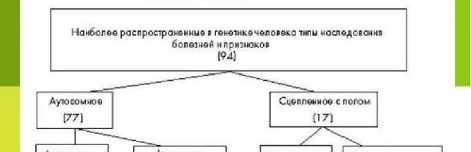
The type of inheritance usually refers to the inheritance of a particular trait depending on whether the gene (allele) that determines it is located on the autosomal or sex chromosome, and whether it is dominant or recessive. In this regard, the following main types of inheritance are distinguished: 1) autosomal dominant, 2) autosomal recessive, 3) sex-linked dominant inheritance and 3) sex-linked recessive inheritance. Of these, 4) sex-limited autosomal and 5) holandric types of inheritance are separately distinguished. In addition, there is 6) mitochondrial inheritance.
At autosomal dominant mode of inheritance the allele of the gene that determines the trait is located in one of the autosomes (non-sex chromosomes) and is dominant. This symptom will appear in all generations. Even when crossing genotypes Aa and aa, it will be observed in half of the offspring.
When autosomal recessive type a trait may not appear in some generations but appear in others. If the parents are heterozygotes (Aa), then they are carriers of a recessive allele, but have a dominant trait. When crossing Aa and Aa, ¾ of the offspring will have a dominant trait and ¼ will have a recessive trait. When crossing Aa and aa in ½, the recessive allele of the gene will manifest itself in half of the descendants.
Autosomal traits occur with equal frequency in both sexes.
Sex-linked dominant inheritance similar to autosomal dominant with one difference: in a sex whose sex chromosomes are the same (for example, XX in many animals is a female organism), the trait will appear twice as often as in a sex with different sex chromosomes (XY). This is due to the fact that if the gene allele is located on the X chromosome of the male body (and the partner does not have such an allele at all), then all daughters will have it, and none of the sons. If the owner of a sex-linked dominant trait is a female organism, then the probability of its transmission is the same to both sexes of descendants.
At sex-linked recessive mode of inheritance Generation skipping may also occur, as in the case of the autosomal recessive type. This is observed when female organisms can be heterozygotes for a given gene, and male organisms do not carry the recessive allele. When a female carrier is crossed with a healthy male, ½ of the sons will express the recessive gene, and ½ of the daughters will be carriers. In humans, hemophilia and color blindness are inherited this way. Fathers never pass the disease gene to their sons (as they only pass on the Y chromosome).
Autosomal, sex-limited mode of inheritance observed when the gene that determines the trait, although localized in the autosome, appears only in one of the sexes. For example, the sign of the amount of protein in milk appears only in females. It is not active in males. Inheritance is approximately the same as in a sex-linked recessive type. However, here the trait can be passed on from father to son.
Hollandic inheritance is associated with the localization of the gene under study on the sex Y chromosome. This trait, regardless of whether it is dominant or recessive, will appear in all sons and not in any daughter.
Mitochondria have their own genome, which determines the presence mitochondrial type of inheritance. Since only the mitochondria of the egg end up in the zygote, mitochondrial inheritance occurs only from mothers (both daughters and sons).
Lecture: Basic types of inheritance of traits in humans
Autosomal dominant the type of inheritance (Fig. 11.2) is characterized by the following features:
1) patients in each generation;
2) a sick child with sick parents;
4) inheritance goes vertically and horizontally;
5) probability of inheritance 100%, 75% and 50%.
It should be emphasized that the above signs of an autosomal dominant type of inheritance will appear only with complete dominance. This is how polydactyly (six-fingered feet), freckles, curly hair, brown eye color, etc. are inherited in humans. With incomplete dominance, hybrids will exhibit an intermediate form of inheritance. If the gene has incomplete penetrance, there may not be patients in every generation.
Autosomal recessive mode of inheritance(Fig. 11.2) is characterized by the following features:
3) men and women are affected equally;
4) inheritance occurs predominantly horizontally;
5) probability of inheritance 25%, 50% and 100%.
Most often, the probability of inheriting a disease of an autosomal recessive type is 25%, since due to the severity of the disease such patients either do not survive to childbearing age,
or do not get married. This is how phenylketonuria, sickle cell anemia, blue eye color, etc. are inherited in humans.
Sex-linked recessive mode of inheritance(Fig. 11.3) is characterized by the following features:
1) patients are not in every generation;
2) healthy parents have a sick child;
3) predominantly men are affected;
4) inheritance occurs mainly horizontally;
5) the probability of inheritance is 25% for all children and 50% for boys.
This is how hemophilia, color blindness, hereditary anemia, muscular dystrophy, etc. are inherited in humans.
Sex-linked dominant mode of inheritance(Fig. 11.4) is similar to autosomal dominant, except that the man passes this trait on to all his daughters (sons receive a Y chromosome from their father, they are healthy). An example of such a disease is a special form of rickets that is resistant to treatment with vitamin B.
Hollandic type of inheritance(Fig. 11.5) is characterized by the following features:
1) patients in all generations;
2) only men get sick;
3) a sick father has all his sons sick;
4) the probability of inheritance is 100% in boys.
Holandric characteristics are not significant in human hereditary pathology. According to the holandric type, men inherit ichthyosis (flaking of the skin), hypertrichosis (excessive hair growth on the ears and external auditory canals), webbing between the toes, etc.
More work on biology
Abstract on biology
Basic types of inheritance of traits
There are many types of inheritance of traits: direct, indirect and complex.
Direct inheritance. in which variants of traits are preserved unchanged from generation to generation - this is the simplest type of inheritance of traits. Direct inheritance is often observed in plants that reproduce vegetatively or produce seeds through self-pollination, less often during the reproduction of animals (within the same breed) or cross-pollination in plants (within the same variety or line).
– Direct inheritance during vegetative propagation of plants
Example 1. Chinatown roses are characterized by bright yellow flowers. When propagated vegetatively, cuttings of this variety always produce plants with bright yellow flowers.
Example 2. Some varieties of weeping willow are characterized by bright yellow shoots. During vegetative propagation, cuttings of these varieties always produce trees with a weeping crown and bright yellow shoots.
– Direct inheritance during self-pollination in plants
Example 1. Pea varieties with green seeds and white flowers, when self-pollinated, always produce green peas in their offspring, from which plants with white flowers grow.
Read also:
Example 2. Tomato varieties with yellow oblong fruits, when self-pollinated, always produce seeds from which plants with yellow oblong fruits grow.
– Direct inheritance during the reproduction of purebred animals and cross-pollination of purebred plants
Example 1. When crossing purebred cows and black-and-white bulls, all their descendants are characterized by a black-and-white color.
Example 2. Cross-pollination of pure tomato plants with red spherical fruits always produces seeds from which plants with red spherical fruits grow.
Indirect inheritance- This is a more complex type of inheritance, which is observed during the reproduction of animals and seed reproduction in plants (which is essentially also sexual). To study indirect inheritance, hybridization is necessary - crossing organisms that differ in genotype. With indirect inheritance, some variants of traits appear in each generation (such traits are called dominant. “dominant”), and other options may temporarily “disappear” and then appear in subsequent generations (such characteristics are called recessive. "retreating")
Example 1. Ancient China is the birthplace of decorative goldfish with a variety of colors, fin lengths and body shapes. Goldfish (as well as carp) are a convenient object for demonstrating crossing: they have external fertilization, and the gametes (spawn and milk) are directly visible. Thousands of years ago, it was noticed that the offspring of dull-colored fish could produce individuals with golden, orange, black and variegated colors. When crossing dull and brightly colored individuals with each other, in some cases all of their offspring had a dull color - this is a dominant trait. However, when these descendants were crossed with each other in subsequent generations, individuals with previously “disappeared” recessive traits reappeared.
Example 2. In medieval Japan, decorative mice with unusual colors were popular: white, yellow, black, spotted. When white and black mice were crossed with each other, in some cases all of their offspring were black (the recessive white color appeared only in subsequent generations), and in other cases - white (now the black color was recessive). Only in the 20th century was it proven that in different cases white color was determined by different genes.
Example 3. When crossing many ornamental plants (snapdragon, night beauty) with red and white flowers, plants with an intermediate pink color grow from hybrid seeds. However, when these pink-flowering hybrid plants are crossed with each other, plants with red, white, and pink flowers appear in their offspring.
Complex types inheritance of traits is called complex because it is very difficult to predict in advance the appearance of new variants of traits. In some cases, new variants of characteristics “suddenly” appear that neither the parents, nor the grandparents, nor the aunts and uncles had. Sometimes such a “sudden” appearance of symptoms is completely unreasonably called a mutation.
Example 1. Aquarium swordtail fish (and a group close to swordtails - platies) are characterized by a variety of colors: greenish-gray, dark red (brick), bright red (scarlet), lemon (light yellow), spotted (tiger and calico). These fish are a convenient subject for demonstration of crossing, since they have internal fertilization, and the females give birth to live fry. When crossing purebred scarlet females with purebred dark red males, greenish-gray hybrids are always obtained. However, when these hybrids are crossed with each other, their offspring produce individuals with a wide variety of colors, including lemon yellow, which all known ancestors did not have.
Example 2. Many food (fruit, berry) and ornamental plants are propagated vegetatively. At the same time, for decades, each variety retains its own characteristics. If you collect seeds from such a plant and sow them, then these seeds will grow into plants with the most fantastic combinations of characteristics.
The type of inheritance is autosomal dominant. Types of inheritance of traits in humans

All the characteristic features of our body are manifested under the influence of genes. Sometimes only one gene is responsible for this, but more often it happens that several units of heredity are responsible for the manifestation of a particular trait.
It has already been scientifically proven that for a person, the manifestation of such characteristics as skin color, hair, eyes, and the degree of mental development depends on the activity of many genes at once. This inheritance does not exactly obey Mendel’s laws, but goes far beyond it.
The study of human genetics is not only interesting, but also important from the point of view of understanding the inheritance of various hereditary diseases. Nowadays, it is becoming quite relevant for young couples to seek genetic counseling so that, after analyzing the pedigree of each spouse, one can confidently say that the child will be born healthy.
Types of inheritance of traits in humans

If you know how a particular trait is inherited, you can predict the likelihood of its manifestation in offspring. All traits in the body can be divided into dominant and recessive. The interaction between them is not that simple, and sometimes it is not enough to know which one belongs to which category.
Now in the scientific world there are the following types of inheritance in humans:
- Monogenic inheritance.
- Polygenic.
- Unconventional.
These types of inheritance, in turn, are also divided into certain varieties.
Monogenic inheritance is based on Mendel's first and second laws. Polygenic is based on the third law. This implies the inheritance of several genes, most often non-allelic.
Non-traditional inheritance does not obey the laws of heredity and is carried out according to its own rules, unknown to anyone.
Monogenic inheritance

This type of inheritance of traits in humans obeys Mendeleev's laws. Considering the fact that there are two alleles of each gene in the genotype, the interaction between the female and male genome is considered separately for each pair.
Based on this, the following types of inheritance are distinguished:
- Autosomal dominant.
- Autosomal recessive.
- X-linked dominant inheritance.
- X-linked recessive.
- Holandric inheritance.
Each type of inheritance has its own characteristics and characteristics.
Signs of autosomal dominant inheritance

The autosomal dominant type of inheritance is the inheritance of predominant traits that are located in autosomes. Their phenotypic manifestations can vary greatly. For some, the symptom may be barely noticeable, but sometimes its manifestation is too intense.
The autosomal dominant type of inheritance has the following characteristics:
- The disease symptom appears in every generation.
- The number of sick and healthy people is approximately the same, their ratio is 1:1.
- If children of sick parents are born healthy, then their children will be healthy.
- The disease affects both boys and girls equally.
- The disease is transmitted equally from men and women.
- The stronger the effect on reproductive functions, the greater the likelihood of various mutations occurring.
- If both parents are sick, then the child, being born homozygous for this trait, is more seriously ill compared to a heterozygote.
All these characteristics are realized only under conditions of complete dominance. In this case, only the presence of one dominant gene will be sufficient for the manifestation of the trait. The autosomal dominant type of inheritance can be observed in humans when inheriting freckles, curly hair, brown eyes and many others.
Autosomal dominant traits

Most people who are carriers of an autosomal dominant pathological trait are heterozygotes for it. Numerous studies confirm that homozygotes for a dominant anomaly have more severe and severe manifestations compared to heterozygotes.
Read also: Contract for the purchase and sale of a car by inheritance - sample
This type of inheritance in humans is characteristic not only of pathological traits, but also some completely normal ones are inherited this way.
Among the normal characteristics with this type of inheritance are:
- Curly hair.
- Dark eyes.
- Straight nose.
- A hump on the bridge of the nose.
- Baldness at an early age in men.
- Right-handedness.
- The ability to roll the tongue into a tube.
- Dimple on the chin.
Among the anomalies that have an autosomal dominant mode of inheritance, the most famous are the following:
- Polyfingered, can be on both hands and feet.
- Fusion of the tissues of the phalanges of the fingers.
- Brachydactyly.
- Marfan syndrome.
- Myopia.
If dominance is incomplete, then the manifestation of the trait cannot be observed in every generation.
Autosomal recessive mode of inheritance

A trait with this type of inheritance can only appear if a homozygote for this pathology is formed. Such diseases are more severe because both alleles of one gene are defective.
The likelihood of manifestation of such signs increases in closely related marriages, therefore in many countries it is prohibited to enter into an alliance between relatives.
The main criteria for such inheritance include the following:
- If both parents are healthy, but are carriers of a pathological gene, then the child will be sick.
- The gender of the unborn child does not play any role in inheritance.
- For one married couple, the risk of having a second child with the same pathology is 25%.
- If you look at the pedigree, you can see a horizontal distribution of patients.
- If both parents are sick, then all children will be born with the same pathology.
- If one parent is sick and the other is a carrier of such a gene, then the probability of having a sick child is 50%
Many metabolic diseases are inherited according to this type.
Type of inheritance linked to the X chromosome

This inheritance can be either dominant or recessive. Signs of dominant inheritance include the following:
- Both sexes can be affected, but women are 2 times more likely.
- If a father is sick, then he can pass on the diseased gene only to his daughters, because the sons receive the Y chromosome from him.
- A sick mother is equally likely to give this disease to children of both sexes.
- The disease is more severe in men because they lack the second X chromosome.
If there is a recessive gene on the X chromosome, then inheritance has the following characteristics:
- A sick child can also be born to phenotypically healthy parents.
- Men are most often affected, and women are carriers of the diseased gene.
- If the father is sick, then you don’t have to worry about the health of your sons; they cannot get a defective gene from him.
- The probability of giving birth to a sick child in a carrier woman is 25%; if we are talking about boys, then it increases to 50%.
This is how diseases such as hemophilia, color blindness, muscular dystrophy, Kallmann syndrome and some others are inherited.
Autosomal dominant diseases

For the manifestation of such diseases, the presence of one defective gene is sufficient, if it is dominant. Autosomal dominant diseases have some characteristics:
- Currently, there are about 4,000 thousand such diseases.
- Both sexes are affected equally.
- Phenotypic demorphism is clearly manifested.
- If a mutation of a dominant gene occurs in gametes, it will most likely appear in the first generation. It has already been proven that men have an increased risk of receiving such mutations with age, which means they can give their children such diseases.
- The disease often manifests itself in all generations.
Inheritance of a defective gene for an autosomal dominant disease has nothing to do with the sex of the child or the degree of development of this disease in the parent.
Autosomal dominant diseases include:
- Marfan syndrome.
- Huntington's disease.
- Neurofibromatosis.
- Tuberous sclerosis.
- Polycystic kidney disease and many others.
All of these diseases can manifest to varying degrees in different patients.
Marfan syndrome

This disease is characterized by damage to connective tissue, and consequently, its functioning. Disproportionately long limbs with thin fingers suggest Marfan syndrome. The type of inheritance of this disease is autosomal dominant.
The following symptoms of this syndrome can be listed:
- Thin build.
- Long "spider" fingers.
- Defects of the cardiovascular system.
- The appearance of stretch marks on the skin for no apparent reason.
- Some patients report pain in muscles and bones.
- Early development of osteoarthritis.
- Rachiocampsis.
- Too flexible joints.
- Possible speech impairment.
- Visual impairment.
You can name the symptoms of this disease for a long time, but most of them are associated with the skeletal system. The final diagnosis will be made after all examinations have been completed and characteristic signs are found in at least three organ systems.
It can be noted that for some, signs of the disease do not appear in childhood, but become obvious somewhat later.
Even now, when the level of medicine is quite high, it is impossible to completely cure Marfan syndrome. Using modern drugs and treatment technologies, it is possible to prolong the life of patients with this disorder and improve its quality.
The most important aspect of treatment is the prevention of the development of aortic aneurysm. Regular consultations with a cardiologist are required. In emergency cases, aortic transplant surgery is indicated.
Huntington's chorea

This disease also has an autosomal dominant mode of inheritance. It begins to appear from the age of 35-50 years. This is due to the progressive death of neurons. Clinically, the following signs can be identified:
- Erratic movements combined with decreased tone.
- Antisocial behavior.
- Apathy and irritability.
- Manifestation of schizophrenic type.
- Mood swings.
Treatment is aimed only at eliminating or reducing symptoms. They use tranquilizers and neuroleptics. No treatment can stop the development of the disease, so death occurs approximately 15-17 years after the first symptoms appear.
Polygenic inheritance

Many signs and diseases have an autosomal dominant mode of inheritance. What this is is already clear, but in most cases it is not so simple. Very often, not one, but several genes are inherited simultaneously. They manifest themselves under specific environmental conditions.
A distinctive feature of this inheritance is the ability to enhance the individual effect of each gene. The main features of such inheritance include the following:
- The more severe the disease, the greater the risk of developing this disease in relatives.
- Many multifactorial traits affect a specific gender.
- The more relatives who have this trait, the higher the risk of developing this disease in future descendants.
All considered types of inheritance belong to the classical variants, but, unfortunately, many signs and diseases cannot be explained because they belong to non-traditional inheritance.
When planning the birth of a baby, do not neglect visiting a genetic consultation. A competent specialist will help you understand your pedigree and assess the risk of having a child with disabilities.
Unforgivable Movie Mistakes You Probably Never Noticed There are probably very few people who don't enjoy watching movies. However, even in the best cinema there are mistakes that the viewer can notice.
15 most beautiful wives of millionaires Check out the list of wives of the most successful people in the world. They are stunning beauties and are often successful in business.
Why have you never seen a baby pigeon? Go to any city square and, without a doubt, you will see hundreds of pigeons flying around passers-by. But, despite such a large number.
AUTOSOMAL DOMINANT TYPE OF INHERITANCE
Examples of diseases: Marfan syndrome, hemoglobinopathy M, Huntington's chorea, colon polyposis, familial hypercholesterolemia, neurofibromatosis, polydactyly.
Autosomal dominant type of inheritance characterized by the following signs:
· Equal frequency of pathology in males and females.
· The presence of patients in each generation of the pedigree, i.e. regular transmission of the disease from generation to generation (the so-called vertical distribution of the disease).
· The probability of having a sick child is 50% (regardless of the gender of the child and the number of births).
· Unaffected family members, as a rule, have healthy offspring (since they do not have the mutant gene).
The listed characteristics are realized under the condition complete dominance(the presence of one dominant gene is sufficient for the development of a specific clinical picture of the disease). This is how freckles, curly hair, brown eye color, etc. are inherited in humans. With incomplete dominance, hybrids will exhibit an intermediate form of inheritance. If the gene has incomplete penetrance, there may not be patients in every generation.
AUTOSOMAL RECESSIVE TYPE OF INHERITANCE
Examples of diseases: phenylketonuria, ocular-cutaneous albinism, sickle cell anemia, adrenogenital syndrome, galactosemia, glycogenosis, hyperlipoproteinemia, cystic fibrosis.
Autosomal recessive mode of inheritance characterized by the following signs:
· Equal frequency of pathology in males and females.
· Manifestation of pathology in the pedigree “horizontally”, often in sibs.
· Absence of the disease in half-blooded (children of the same father from different mothers) and half-brothers (children of the same mother from different fathers).
· The patient's parents are usually healthy. The same disease can be detected in other relatives, for example, in cousins or second cousins of the patient.
The appearance of autosomal recessive pathology is more likely in consanguineous marriages due to the greater likelihood of meeting two spouses who are heterozygous for the same pathological allele received from their common ancestor. The greater the degree of relationship between the spouses, the higher this probability. Most often, the probability of inheriting a disease of an autosomal recessive type is 25%, since due to the severity of the disease, such patients either do not live to childbearing age or do not marry.
CHROMOSOME-LINKED X-DOMINANT INHERITANCE
Examples of diseases: one form of hypophosphatemia is vitamin D-resistant rickets; Charcot-Marie-Tooth disease - linked dominant; Orofacial-digital syndrome type I.
Signs of the disease:
· Males and females are affected, but women are 2 times more likely.
· Transfer of a pathological allele by a sick man to all daughters and only to daughters, but not to sons. Sons receive the Y chromosome from their father.
· Transmission of the disease by a sick woman to both sons and daughters is equally likely.
· The disease is more severe in men than in women.
CHROMOSOME-LINKED X-RECESSIVE INHERITANCE
Examples of diseases: hemophilia A, hemophilia B; X-linked recessive Charcot-Marie-Tooth disease; color blindness; Duchenne-Becker muscular dystrophy; Kallmann syndrome; Hunter's disease (mucopolysaccharidosis type II); hypogammaglobulinemia of Brutonian type.
Signs of the disease:
· Patients are born to phenotypically healthy parents.
· The disease occurs almost exclusively in males. Mothers of patients are obligate carriers of the pathological gene.
· A son never inherits a disease from his father.
· A carrier of a mutant gene has a 25% chance of having a sick child (regardless of the sex of the newborn); the probability of having a sick boy is 50%.
HOLANDRIC, OR LINKED TO CHROMOSOME Y,
TYPE OF INHERITANCE
Examples of signs: ichthyosis of the skin, hypertrichosis of the ears, excess hair growth on the middle phalanges of the fingers, azoospermia.
Signs:
· Transfer of a trait from the father to all sons and only sons.
· Daughters never inherit a trait from their father.
· “Vertical” nature of inheritance of a trait.
· The probability of inheritance for males is 100%.
MITOCHONDRIAL INHERITANCE
Examples of diseases(mitochondrial diseases): Leber optic atrophy, Leigh syndrome (mitochondrial myoencephalopathy), MERRF (myoclonic epilepsy), familial dilated cardiomyopathy.
Signs of the disease:
· The presence of pathology in all children of a sick mother.
· The birth of healthy children from a sick father and a healthy mother.
These features are explained by the fact that mitochondria are inherited from the mother. The portion of the paternal mitochondrial genome in the zygote is DNA from 0 to 4 mitochondria, and the maternal genome is DNA from approximately 2500 mitochondria. In addition, it appears that after fertilization, paternal DNA replication is blocked.
With all the diversity of gene diseases in their pathogenesis there is general pattern: the beginning of the pathogenesis of any gene disease is associated with primary effect of the mutant allele- a pathological primary product (qualitatively or quantitatively), which is included in the chain of biochemical processes and leads to the formation of defects in the cellular, organ And organismal levels.
Pathogenesis of the disease at the molecular level unfolds depending on the nature of the mutant gene product in the form of the following disorders:
Abnormal protein synthesis;
Lack of primary product production (most common);
Production of a reduced amount of normal primary product (in this case, the pathogenesis is highly variable);
Production of an excess amount of product (this option is only assumed, but has not yet been discovered in specific forms of hereditary diseases).
Options for implementing the action of an abnormal gene:
1) abnormal gene → cessation of mRNA synthesis → cessation of protein synthesis → hereditary disease;
2) abnormal gene → cessation of mRNA synthesis → hereditary disease;
3) an abnormal gene with a pathological code → synthesis of pathological mRNA → synthesis of pathological protein → hereditary disease;
4) disruption of switching genes on and off (repression and depression of genes);
5) abnormal gene → lack of hormone receptor synthesis → hereditary hormonal pathology.
Examples of the 1st variant of gene pathology: hypoalbuminemia, afibrinogenemia, hemophilia A (factor VIII), hemophilia B (IX - Christmas factor), hemophilia C (XI factor - Rosenthal), agammaglobulinemia.
Examples of the 2nd option: albinism (enzyme deficiency - tyrosinase → depigmentation); phenylketonuria (phenylalanine hydroxylase deficiency → phenylalanine accumulates → its metabolic product, phenylpyruvate, is toxic to the central nervous system → mental retardation develops); alkaptonuria (deficiency of homogentisic acid oxidase → homogentisic acid accumulates in the blood, urine, tissues → coloring of tissues, cartilage); enzymopathic methemoglobinemia (methemoglobin reductase deficiency → methemoglobin accumulates → hypoxia develops); adrenogenital syndrome (one of the most common hereditary human diseases: frequency in Europe 1:5000, among Eskimos of Alaska 1:400 - 1:150; 21-hydroxylase defect → cortisol deficiency, accumulation of androgens → in men - accelerated sexual development, in women - virilization).
Example of the 3rd variant of gene pathology: M - hemoglobinosis (abnormal M-hemoglobin is synthesized, which differs from normal A-hemoglobin in that at position 58 of the α-chain (or at position 63 of the β-chain) histidine is replaced by tyrosine → M-hemoglobin enters into a strong bond with oxygen, not giving it to the tissues, it forms methemoglobin → hypoxia develops).
Example of option 4: Thalassemia. It is known that fetal red blood cells contain special fetal hemoglobin, the synthesis of which is controlled by two genes. After birth, the action of one of these genes is inhibited and another gene is turned on, providing the synthesis of Hb A (95-98% of hemoglobin in healthy people). In pathology, persistence of fetal hemoglobin synthesis may be observed (its amount in healthy people is 1-2%). Hb S is less stable than Hb A - therefore hemolytic anemia develops.
Example of option 5: testicular feminization. It has been revealed that individuals with this disease do not have testosterone receptors. Therefore, the male embryo acquires features characteristic of the female body.
Pathogenesis of any hereditary disease in different individuals, although similar in primary mechanisms and stages, is formed strictly individually– the pathological process, launched by the primary effect of the mutant allele, acquires integrity with natural individual variations depending on the genotype of the organism and environmental conditions.
Characteristics clinical picture gene diseases are caused by the principles expression, repression and gene interaction.
The following are distinguished: main characteristics of gene diseases:features of the clinical picture; clinical polymorphism; genetic heterogeneity. At the same time, it is impossible to fully observe all the common features in one disease. Knowledge of the general features of gene diseases will allow the doctor to suspect a hereditary disease even in a sporadic case.
Features of the clinical picture:
variety of manifestations- the pathological process affects several organs already at the initial stages of the formation of the disease;
different ages of onset of the disease;
progression of the clinical picture and chronic course;
Conditioned disability since childhood and shortened life expectancy.
The variety of manifestations and the involvement of many organs and tissues in the pathological process for this group of diseases is due to the fact that the primary defect is localized in the cellular and intercellular structures of many organs. For example, in hereditary connective tissue diseases, the synthesis of a protein of one or another fibrous structure specific to each disease is disrupted. Since connective tissue is present in all organs and tissues, the variety of clinical symptoms in these diseases is a consequence of connective tissue abnormalities in various organs.
Age of onset for this group of diseases practically unlimited: from the early stages of embryonic development (congenital defects) – to old age ( Alzheimer's disease). The biological basis of different ages of onset of gene diseases lies in the strictly temporal patterns of ontogenetic regulation of gene expression. The reasons for different ages of onset of the same disease may be the individual characteristics of the patient’s genome. The effect of other genes on the manifestation of the effect of the mutant gene may change the time of development of the disease. The time of onset of the action of pathological genes and environmental conditions are also important, especially in the prenatal period. Generalized data on the timing of clinical manifestation of gene diseases indicate that 25% of all gene diseases develop in utero, and almost 50% of gene diseases manifest themselves in the first three years of life.
Most gene diseases are characterized by progression of the clinical picture And chronic protracted course with relapses. The severity of the disease “increases” as the pathological process develops. Primary biological basis This characteristic is the continuity of functioning of the pathological gene (or the absence of its product). This is accompanied by enhancing the pathological process secondary processes: inflammation; dystrophy; metabolic disorders; hyperplasia.
Most gene diseases are severe and lead to disability in childhood And shortens life expectancy. The more important a monogenically determined process is in ensuring life activity, the more clinically severe the manifestation of the mutation.
Concept "clinical polymorphism" unite:
Variability: timing of the onset of the disease; severity of symptoms; duration of the same illness;
Tolerance to therapy.
The genetic causes of clinical polymorphism can be determined not only by the pathological gene, but also by the genotype as a whole, that is, the genotypic environment in the form of modifier genes. The genome as a whole functions as a well-coordinated system. Together with the pathological gene, the individual inherits from his parents combinations of other genes that can enhance or weaken the effect of the pathological gene. In addition, in the development of a gene disease, like any hereditary trait, not only the genotype, but also the external environment matters. There is a lot of evidence for this position from clinical practice. For example, the symptoms of phenylketonuria in a child are more severe if during its prenatal development the mother’s diet included a lot of foods rich in phenylalanine.
There is a concept genetic heterogeneity masquerading as clinical polymorphism.
Genetic heterogeneity means that the clinical form of a gene disease can be caused by:
mutations in different genes, encoding enzymes of one metabolic pathway;
different mutations in the same gene, leading to the emergence of different alleles (multiple alleles).
In fact, in these cases we are talking about different nosological forms, from an etiological point of view, combined into one form due to the clinical similarity of the phenotype. The phenomenon of genetic heterogeneity is general; it can be called a rule, since it applies to all proteins of the body, including not only pathological, but also normal variants.
Deciphering the heterogeneity of gene diseases continues intensively in two directions:
clinical- the more accurately studied phenotype(analysis of the clinical picture of the disease), the more opportunities there are in the discovery of new forms of diseases, in the division of the studied form into several nosological units;
genetic- provides the most complete information about the heterogeneity of the clinical form of the disease DNA probe method(a modern method for analyzing human genes). The assignment of a gene to one or different linkage groups, the localization of the gene, its structure, the essence of the mutation - all this makes it possible to identify nosological forms.
Concept genetic heterogeneity of gene diseases opens up many opportunities in understanding the essence of individual forms and causes of clinical polymorphism, which is extremely important for practical medicine and provides the following opportunities: correct diagnosis; choice of treatment method; medical and genetic counseling.
Understanding epidemiology of gene diseases necessary for a doctor of any specialty, since in his practice he may encounter manifestations of a rare hereditary disease within the area or population he serves. Knowledge of the patterns and mechanism of spread of gene diseases will help the doctor timely develop preventive measures: examination for heterogeneity; genetic counseling.
Epidemiology of gene diseases includes the following information:
About the prevalence of these diseases;
About the frequencies of heterozygous carriage and the factors that determine them.
Prevalence of the disease(or number of patients) in the population determined by population patterns: the intensity of the mutation process; selection pressure, which determines the fertility of mutants and heterozygotes under specific environmental conditions; population migration; isolation; genetic drift. Data on the prevalence of hereditary diseases are still fragmentary due to the following reasons: a large number of nosological forms of genetic diseases; their rarity; incomplete clinical and pathological diagnosis of hereditary pathology. The most objective assessment of the prevalence of these diseases in different populations is to determine their number among newborns, including stillborns. The overall frequency of newborns with genetic diseases in populations as a whole is approximately 1%, of which:
With an autosomal dominant type of inheritance - 0.5%;
With autosomal recessive - 0.25%;
X-linked - 0.25%;
Y-linked and mitochondrial diseases are extremely rare.
The prevalence of individual forms of disease ranges from 1:500 (primary hemochromatosis) up to 1:100000 and below (hepatolenticular degeneration, phenylketonuria).
The prevalence of a gene disease is considered:
High – if 1 patient occurs per 10,000 newborns or more often;
Basic symbols:
When constructing pedigrees, it is necessary to observe following rules :
a) it is necessary to find out the number of generations from the collected history;
b) the pedigree begins to be built from the proband;
c) each generation is numbered with Roman numerals on the left;
d) symbols indicating individuals of one generation are located on a horizontal line and numbered in Arabic numerals.
There are the following types of inheritance signs:
autosomal dominant;
autosomal recessive;
X-linked (sex-linked) dominant;
X-linked (sex-linked) recessive;
holandric.
Autosomal dominant type of inheritance
A sick child is born to sick parents with a 100% probability if they are homozygous; 75% if they are heterozygous.
Autosomal recessive mode of inheritance
Both men and women are affected equally.
The probability of having a sick child from healthy parents is 25% if they are heterozygous, 0% if both of them or one of them is homozygous for the dominant gene.
Often manifests itself in consanguineous marriages.
X-linked (sex-linked) dominant type of inheritance
Sick people occur in every generation.
Women are more affected.
If a father is sick, then all his daughters are sick.
A sick child is born to sick parents with a probability of 100% if the mother is homozygous, 75% if the mother is heterozygous.
The probability of having a sick child from healthy parents is 0%.
X-linked (sex-linked) recessive mode of inheritance
Patients do not occur in every generation.
Mostly men are affected.
The probability of having a sick boy born to healthy parents is 25%, and a sick girl is 0%.
Hollandic type of inheritance
Sick people occur in every generation.
Only men get sick.
If a father is sick, then all his sons are sick.
The probability of having a sick boy from a sick father is 100%.
Twin method (proposed in 1876 by F. Galton) is the study of genetic patterns in twins.
The essence of the method: comparison of characteristics in different groups of twins based on their similarities (concordance) or differences (discordance).
Method steps:
1. Compiling a sample of twins from the entire population.
2. Diagnosis of zygosity of twins.
3. Establishing the relative role of heredity and environment in the formation of a trait.
D  To assess the role of heredity and environment in the formation and development of a trait, they use Holzinger's formula:
where N is the proportion of hereditary factors, KMB% and KDB% concordance of monozygotic and dizygotic twins as a percentage.
If H is greater than 0.5, then the genotype plays a large role in the formation of the trait; if H is less than 0.5, then the environment plays a large role.
Cytogenetic method - This is the study of a karyotype using microscopic technology.
Method steps:
1. Obtaining and culturing cells (lymphocytes, fibroblasts) on artificial nutrient media.
2. Addition of phytohemagglutinin to the nutrient medium to stimulate cell division.
3. Stopping cell division at the metaphase stage by adding colchicine.
4. Treatment of cells with a hypotonic NaCl solution, as a result of which the cell membrane is destroyed and a “scattering” of chromosomes is obtained.
5. Staining of chromosomes with specific dyes.
6. Microscoping and photographing chromosomes.
7. Drawing up an idiogram and its analysis.
The method allows:
a) diagnose genomic and chromosomal mutations;
b) determine the genetic sex of the organism.
Population statistical method - is the study of the genetic composition of a population. It is based on the Hardy-Weinberg law and allows us to determine the frequency of genes and genotypes in different populations (see section VII).
Biochemical methods. The cause of most hereditary monogenic diseases are metabolic defects associated with enzymopathies (disturbances in the structure of enzymes involved in metabolic reactions). At the same time, intermediate metabolic products accumulate in the body, and therefore, by determining them or the activity of enzymes, using biochemical methods it is possible to diagnose hereditary metabolic diseases (gene mutations). Quantitative biochemical methods (stress tests) make it possible to identify heterozygous carriage of a pathological recessive gene.
Recombinant DNA Methods(DNA cloning and nucleic acid hybridization)- allow you to detect a pathological gene in the genome if this gene is sequenced (the nucleotide sequence is completely determined).
Dermatoglyphic analysis is the study of human ridge skin (skin of the fingertips, palmar side of the hands and plantar side of the feet), where the papillary layer of the dermis is strongly pronounced.
The method is applied:
a) to establish the zygosity of twins;
b) as an express method for diagnosing the congenital component of some hereditary diseases.
Typically, with genomic pathology, a combination of certain indicators is noted: radial loops on the 4th and 5th fingers, four-digit groove, main palmar angle from 60 0 to 80 0, etc.
Chemical methods based on high-quality color chemical reactions. They are used for preliminary diagnosis of hereditary metabolic diseases. As a screening test diagnosis of phenylketonuria The method of wetting strips of paper soaked in a 10% solution of FeCl 3 or 2,4 dinitrophenylhydrazine with the child’s urine is used. If phenylpyruvic acid is present in the urine, a greenish color appears on the filter paper.
Definition of X- andY-sex chromatin. Buccal epithelial cells or leukocytes are used for research. X-chromatin is determined by staining the preparation acetorcein, and Y-chromatin - when stained Acrychinipriet. These methods make it possible to identify the number of sex chromosomes in a karyotype (the number of X chromosomes is always one more than the number of X-chromatin clumps, the number of Y chromosomes is equal to the number of Y-chromatin clumps); establish the genetic sex of an individual, diagnose chromosomal diseases of sex (in combination with other methods).
Prenatal diagnostic methods make it possible to identify hereditary defects of the fetus in the early stages of pregnancy.
1. Indirect methods (the object of the study is a pregnant woman).
2. Direct methods ( the object of study is the fetus).
Indirect methods are indicated for all pregnant women and include:
obstetric and gynecological examination;
medical genetic examination;
serological examination;
study of the content of embryo-specific proteins.
Level -FP is increased with: threatened miscarriage; intrauterine fetal death; Rh incompatibility between mother and fetus; multiple pregnancy; umbilical cord hernia; neural tube defects; sacrococcygeal teratoma. Low level -FP observed in Down syndrome.
Direct research methods: non-invasive (ultrasonography) andinvasive (amniocentesis, chorionic villus biopsy, fetoscopy).
Direct non-invasive methods are indicated to all pregnant women women (safe for mother and fetus).
Ultrasonography - the use of ultrasound to obtain an image of the fetus and its membranes on a monitor. The method is used at 12–15 weeks of pregnancy, when all organ systems of the fetus are formed, and makes it possible to identify multiple pregnancies and major defects (hydrocephalus, anencephaly, aplasia of the limbs, etc.).
Direct invasive methods are carried out according to strict indications (woman’s age is more than 35 years; structural rearrangements of chromosomes in one of the parents; the first child has a disease with a recessive type of inheritance; pregnancy due to psychological trauma, serious illness, etc.).
Amniocentesis. After treating the skin, the abdominal wall and uterus are pierced with a needle, and amniotic fluid is extracted with a syringe under the control of ultrasonography. The liquid is used for biochemical research (diagnosis of gene mutations), and the fetal cells contained in it are used for cytogenetic research (diagnosis of chromosomal and genomic mutations).
Chorionic villus biopsy performed under ultrasonography control at 8–12 weeks of pregnancy. Using a catheter with a plastic cannula connected to a syringe, 10–25 mg of chorionic villi are extracted and used for cytogenetic, biochemical studies and DNA analysis. Method allows you to diagnose various mutations in early pregnancy.
Fetoscopy (amnioscopy) carried out at 18–22 weeks of pregnancy using a fiberoptic endoscope inserted under local anesthesia into the amnion cavity through the abdominal wall of the uterus. The method allows examine the fetus, placenta, umbilical cord and take fetal blood from the umbilical cord to diagnose hereditary blood diseases. Rarely used; in 5–10% of cases it can cause miscarriage.